


Understanding Radiation: Comparing X-ray, CT, and Mammography
Learn the difference between X-ray, CT, and mammography scans. Understand how each test uses radiation, what they are used for, and how safe they are in everyday medical care.
Radiology plays an important role in the early detection and diagnosis of many health conditions. When a doctor refers someone for a scan, they are often choosing between different imaging methods. Three of the most commonly used types of scans that involve radiation are X-ray, CT (computed tomography), and mammography. While they all use radiation to create images of the body, the way they work and what they are best suited for can be very different.
This article will explain what each of these tests is, how they use radiation, and when each might be used. Understanding these differences helps patients feel more informed and calm when it comes time for a scan.
What Is Radiation in Medical Imaging?
Radiation in medical imaging refers to ionising radiation, which has enough energy to remove electrons from atoms. This type of radiation can pass through the body and form images of internal structures. While it sounds concerning, the levels used in medical scans are carefully managed and kept low.
The main purpose of using radiation in scans is to get a clear look at bones, organs, or soft tissues that cannot be seen from the outside. Doctors then use these images to help make decisions about a persons health, such as finding broken bones, checking for disease, or spotting changes in the body.
How an X-ray Works
An X-ray is often the first type of scan someone may have in a medical setting. It uses a small amount of ionising radiation to take a snapshot of the inside of the body. When the radiation passes through the body, it is absorbed in different amounts by different tissues. Dense materials like bones absorb more radiation and appear white on the X-ray image. Softer tissues appear in shades of grey.
Common Uses
-
Identifying broken bones
-
Detecting infections like pneumonia in the lungs
-
Looking for arthritis in joints
-
Locating dental problems
Radiation Dose
The radiation from a single chest X-ray is about 0.1 millisieverts (mSv). To put this into context, the average person in Australia receives around 1.5 to 2.0 mSv per year from natural sources like soil and sunlight. A chest X-ray is equivalent to about 10 days of natural background radiation.
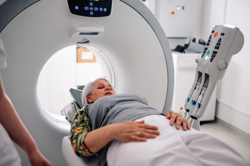
How a CT Scan Works
CT, short for computed tomography, uses X-rays taken from many different angles. These are then processed by a computer to create detailed cross-sectional images of the body. While X-rays give flat, two-dimensional images, CT scans provide a more complete and layered view.
CT scans are helpful for examining both bone and soft tissue in great detail. They are often used when a more thorough look is needed or when symptoms are not clear.
Common Uses
-
Checking for internal injuries after an accident
-
Diagnosing cancers or monitoring tumour growth
-
Investigating causes of chest or abdominal pain
-
Planning surgeries and other medical treatments
Radiation Dose
Because CT scans take many X-ray images, the dose is usually higher than a regular X-ray. A typical CT scan of the chest might be 7 mSv, which is around the same radiation as two years of natural exposure. The dose can vary depending on which part of the body is being scanned and how detailed the images need to be.
How Mammography Works
Mammography is a specialised form of X-ray used to examine the breast tissue. It can detect changes in the breast, such as lumps or unusual patterns, often before they can be felt by hand. This makes it a valuable tool for detecting breast cancer early.
There are two main types of mammograms:
-
Screening mammograms, which are used when there are no symptoms
-
Diagnostic mammograms, which are used when a lump or other concern has been found
Common Uses
-
Early detection of breast cancer
-
Investigating lumps, pain, or nipple changes
-
Monitoring changes over time in breast tissue
Radiation Dose
A typical mammogram exposes a person to 0.4 mSv, which is similar to seven weeks of natural background radiation. Although the dose is higher than a chest X-ray, it is still within safe limits, especially considering the potential to find cancer at an early and treatable stage.
Comparing X-ray, CT, and Mammography
|
Scan Type |
Image Detail |
Radiation Dose |
Common Uses |
|
X-ray |
Basic |
Low (0.1 mSv) |
Bones, chest, joints |
|
CT |
High (cross-sectional) |
Moderate to High (210 mSv) |
Organs, trauma, cancer |
|
Mammography |
Moderate (specialised) |
Low to Moderate (0.4 mSv) |
Breast tissue |
It is important to remember that these doses are well below harmful levels and are used only when necessary. The medical benefit of accurate diagnosis far outweighs the small risk from radiation.
Why This Knowledge Matters
Many people worry when they hear the word radiation. It is a natural reaction. However, understanding how these scans work and the safety steps taken by medical staff can make the experience much less stressful. Each scan is ordered for a specific reason. Knowing the purpose helps patients feel more comfortable and confident during the process.
In some cases, people may be referred for follow-up scans or additional imaging. This does not always mean something is wrong. It might simply be a way to get more information or to confirm results.
Local Access to Trusted Imaging Services
In Queensland, patients have access to clinics that focus on clarity, care, and accurate diagnosis. For example, some radiology providers in southeast Queensland offer a range of imaging services such as X-ray, CT Scan, and mammography. These services are often available by appointment and some are bulk billed, depending on the type of scan and referral. Their teams work with general practitioners and specialists to ensure timely results, especially when early diagnosis is important. This helps people get the answers they need without long delays or unnecessary worry.
Final Thoughts
X-ray, CT, and mammography are valuable tools in medical care. Each uses radiation in a different way to help doctors see inside the body and make important decisions about health. While the idea of radiation can be unsettling at first, the actual doses used in these scans are small and carefully controlled.
By learning about the purpose and safety of each scan, patients can take part in their care with more confidence. Whether someone needs a quick check of a sore knee, a deeper look into unexplained pain, or an early check for breast cancer, these imaging tools play a key role in keeping people well-informed and on the path to good health.